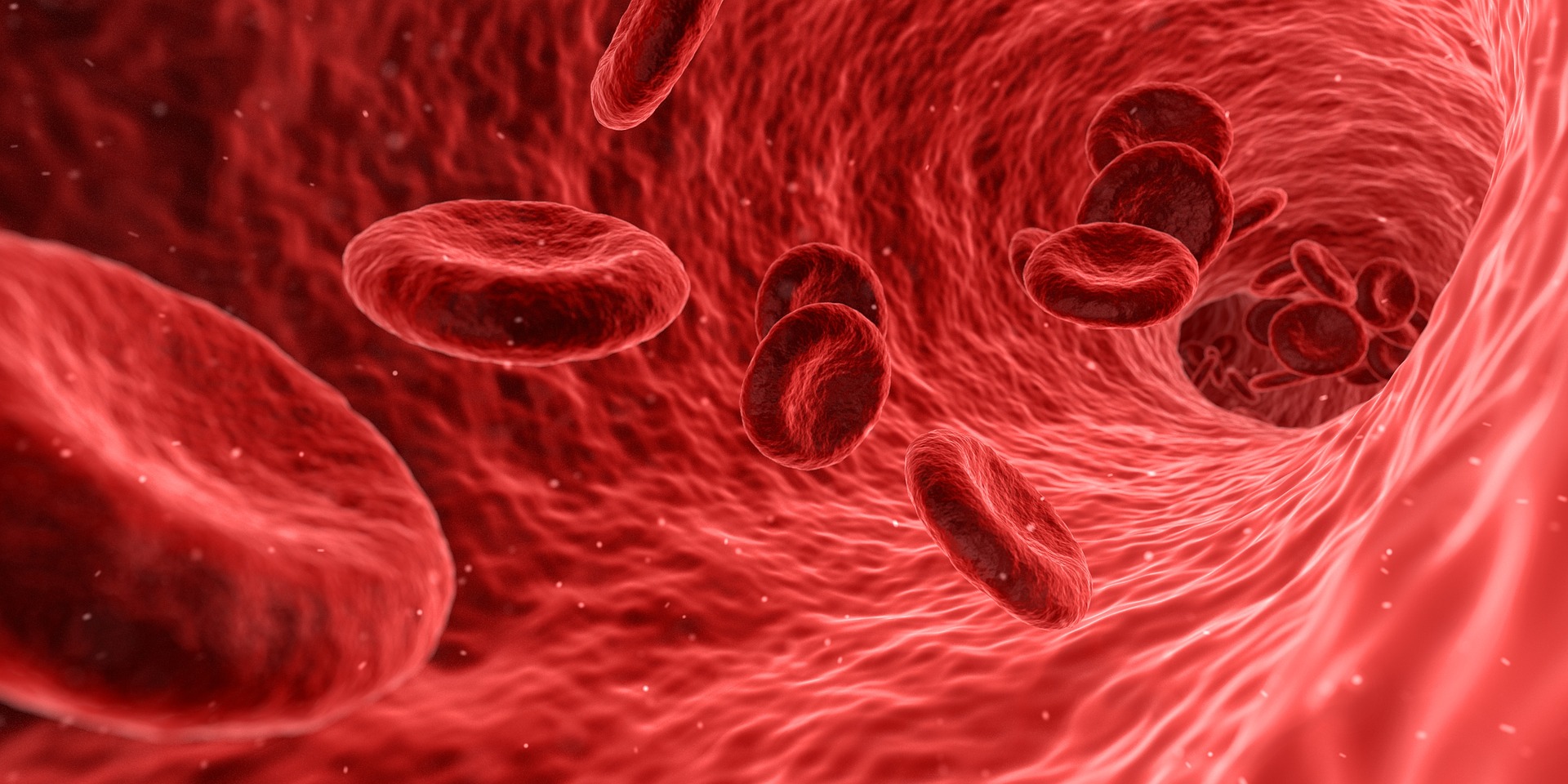
 The circulatory system delivers nutrients wherever they are needed by using the heart to pump the blood out to the cells.
Arteries are any of the tubular, muscular and elastic walled vessels that carry blood from the heart through the body. They are said to “branch,” “diverge,” or “fork” as they form smaller and smaller divisions. Veins on the other hand, carry blood towards the heart. They are said to “join,” “merge” and “converge” into successively larger vessels as they approach the heart. Capillaries are the smallest of the blood vessels and the sites of exchange between the blood and tissue cells.
Arteries: Bringing In The Nutrients:

Numerous arteries bring blood to the digestive system. Since digestion begins in the mouth, the arteries of that area are worth mentioning. The external carotid arteries supply most of the tissue of the head with blood with the exception of the brain and orbits. This artery sends branches to the thyroid gland and larynx, and to the tongue (lingual artery). It ends by splitting into the superficial temporal artery, which supplies the parotid salivary gland, and the maxillary artery, which supplies the upper and lower jaws and chewing muscles, the teeth and the nasal cavity.
The esophagus has a few different arteries that supply blood to it such as the esophageal branch of the inferior thyroid artery (right side), esophageal branch of the inferior thyroid artery (left side), esophageal branch of thoracic aorta, and the esophageal branch of the left gastric artery.
Directly below the diaphragm, the Celiac Trunk, a very large unpaired branch of the abdominal aorta (artery) somewhere around T12 (the twelfth thoracic vertebra) divides almost immediately into three branches: the common hepatic, splenic and the left gastric arteries. The common hepatic artery gives off branches to the stomach, duodenum and the pancreas. The branch serving the duodenum, the gastroduodenal artery also branches off and becomes the hepatic artery proper, which splits into right and left branches that serve the liver. The splenic artery passes deep into the stomach and sends branches to the pancreas and stomach and terminates in branches to the spleen.
A short note on mesenteries. Mesenteries are double-layered extensions of the peritoneum that support most organs in the abdominal cavity. Peritoneum is serous membrane (a thin double-layered membrane that folds in on itself). The parietal serosa lines the cavity walls of the ventral body cavity (which contains both the thoracic cavity and the abdominopelvic cavity), and the visceral serosa, covers the organs in the cavity. The serous membranes are separated by a thin layer of lubricating fluid called serous fluid, which is secreted by both the membranes. It is this fluid that allows the organs to move around enough but not too much lending to their efficiency.
The Superior Mesenteric artery is a large unpaired artery arising from the abdominal aorta at the L1 level (the first lumbar vertebra). It runs to the pancreas and then to the mesentery where branches serve the small intestine via the intestinal arteries and most of the large intestine, the appendix, cecum, ascending colon, and part of the transverse colon.
The Inferior Mesenteric artery is the final branch of the abdominal aorta and is unpaired and arises from the anterior aortic surface at the L3 level (third lumbar vertebra). It serves the distal part of the large intestine (away from the origin), from the midpart of the transverse colon to the midrectum.
Another amazing feature that these two arteries, the superior mesenteric artery and the inferior mesenteric artery have is that looping anastomoses (communication) between the superior and the inferior arteries help ensure that blood will continue to reach the digestive system in cases of trauma to one of these abdominal arteries. Remember that this area is not surrounded by bone like the thoracic cavity and therefore is vulnerable in situations like car accidents.
Veins- Blood leaving the digestive system carrying the nutrients:

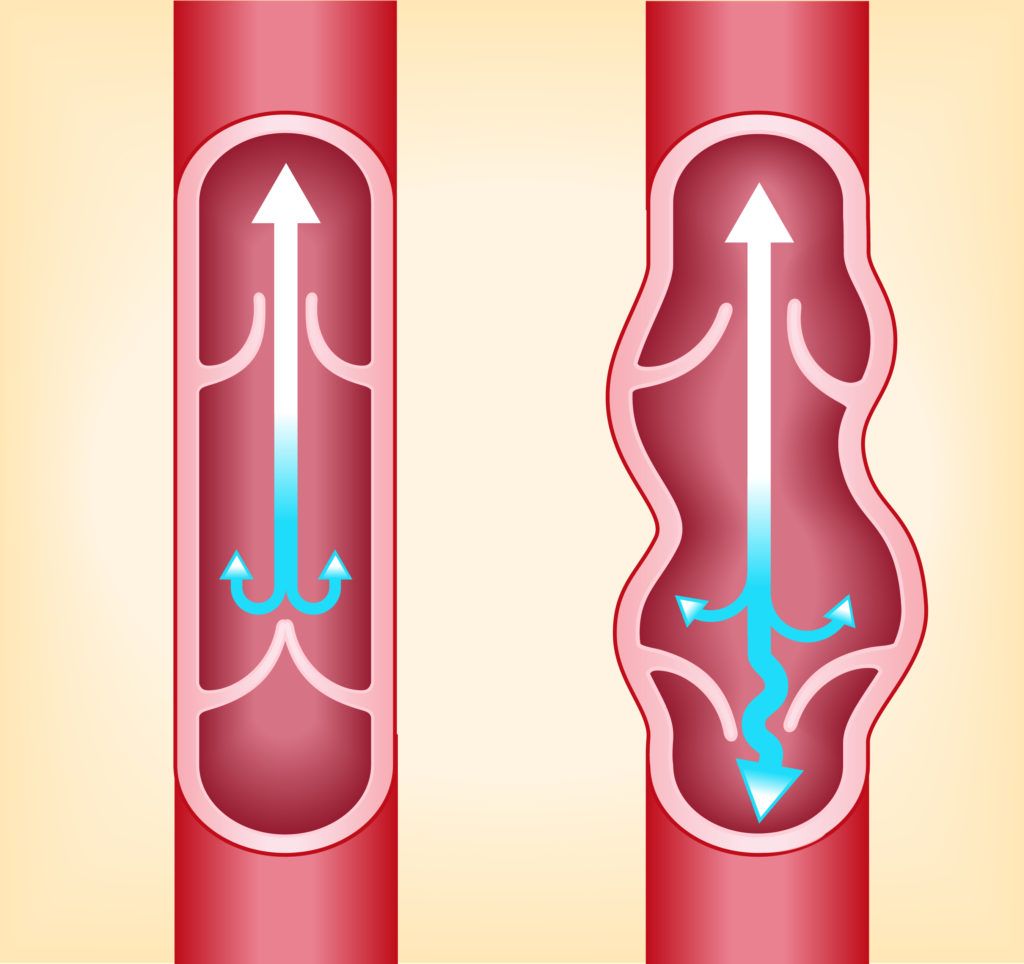
Veins are vessels that carry blood from the capillaries toward the heart and have thinner walls than arteries. They usually have some valves at intervals to prevent reflux. (left side of the picture shows valves working correctly and vein on the right side of the picture shows blood reflux). Blood moving back to the heart has less hemoglobin (an iron containing respiratory pigment) and therefore is dark-colored.
Many illustrations will show veins as “blue” vessels. This is only to designate that they are veins and not arteries. Veins are red in color. Sometimes even the veins look blue through your skin, but that is just the pigments in your skin that cause them to “appear” blue.
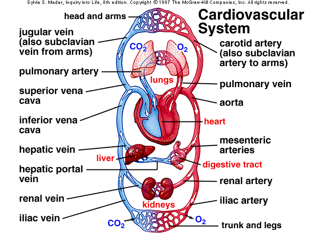
Unlike most veins that deliver the blood directly towards the heart, there are two exceptions: First the venous blood draining from the brain enters large sinuses rather than typical veins and second, blood draining from the digestion organs enters a special sub-circulation, the hepatic portal circulation and passes through the liver before it reenters the general systemic circulation.
The hepatic portal vein directs the blood leaving the digestive system to the liver. Every cell in the liver has access to this blood, via the capillaries that branch off from this vein. The blood leaving the liver then again collects into a vein called the hepatic vein which returns blood to the heart.
The liver, which is the body’s most important metabolic organ, performs many jobs in preparing the absorbed nutrients for use by the body. The anatomical placement of the liver in the body ensures that it will be the first to receive the nutrients absorbed from the GI tract. The liver also helps protect the brain and heart from ingested poisons or toxins that get past the intestinal cells. When this happens, the liver itself may become damaged. (A few examples being viruses such as hepatitis, drugs such as barbiturates, or alcohol, and toxins such a pesticide residues.)
Another way nutrients travel is though lymph. Lymph is clear yellowish fluid that is similar to blood except that it contains no red blood cells or platelets. This fluid comes from the spaces between the tissues and it is thought that it is muscle contractions that creates this liquid. The system that lymph travels in is called the lymphatic system. Unlike the circulatory system which uses a pump (the heart), the lymphatic system has no pump. Muscle contractions and pressure created by those contractions move lymph from one part of the body to another. Lymph from the GI tract transports fat and fat-soluble vitamins to the bloodstream via the lymphatic vessels. The lymph eventually collects behind the heart in the thoracic duct. The duct opens into the subclavian vein, where the lymph enters the bloodstream. One fact that should be noted: Nutrients that enter the bloodstream through the lymph have never gone through the liver. They have however, traveled through lymph nodes. There are hundreds of lymph nodes (small organs filled with macrophages-protective cells which attack microorganisms and other toxins) throughout the body, and also clustered in the groin, underarm region, and neck area. They serve two major functions, both protecting the body. First they act as filters (macrophages), cleaning out the lymph and second they help to activate the immune system.
A General View of How Blood Flows Through the System:

- Blood leaves the right side of the heart by way of the pulmonary artery (an arterial trunk or either of its two main branches that carry oxygen-deficient blood to the lungs). The right side of heart deals with carbon dioxide.
- Blood loses carbon dioxide and picks up oxygen in the lungs and returns to the left side of the heart by way of the pulmonary vein. Left side of brain deals with oxygenated blood.
- Blood leaves the left side of the heart by way of the aorta, the main artery that launches blood on its course through the body.
- Blood may leave the aorta to go to the upper body and heart; or blood may leave the aorta to go to the lower body.
- Blood may go to the digestive tract and then the liver or blood may go to the pelvis, kidneys, and legs.
- Blood returns to the right side to the heart. Lymph from most of the body’s organs, including the digestive system , enters the bloodstream through the thoracic duct behind the heart which opens into the subclavian vein, where the lymph enters the bloodstream.
- Once inside the vascular system, the nutrients can travel freely to any destination and can be taken into the cells and used as needed.
